
Discussion
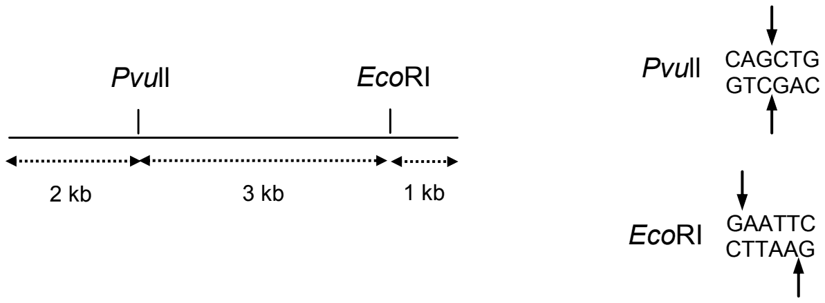
The figure below is a schematic of a linear double stranded DNA containing the indicated restriction sites. The DNA was completely digested with PvuII and EcoRI. The products were purified and added to an appropriately buffered reaction mixture containing dNTP mix, α-32P dATP, and Klenow fragment of E. coli DNA polymerase I. The Klenow reaction products were analyzed by gel electrophoresis and autoradiography. Which of the following products depicts the expected result?
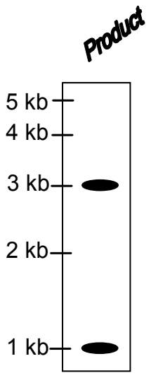
- A.
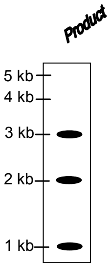
- B.
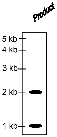
- C.
- D.
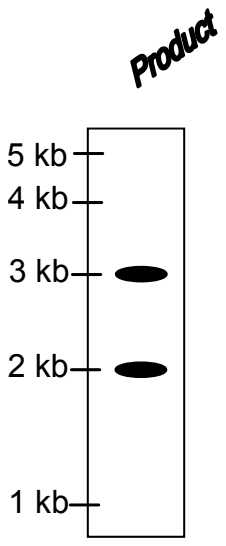
Answer: A
The expected result is (A): labelled bands at 3 kb and 1 kb only.
Digestion gives three fragments: 2 kb (left end to PvuII), 3 kb (PvuII to EcoRI) and 1 kb (EcoRI to right end). PvuII cuts CAG/CTG in the middle, leaving blunt ends. EcoRI cuts G/AATTC, leaving a 5' overhang of AATT with a recessed 3' end opposite it.
Klenow fragment fills in a recessed 3' end using the overhang as template; filling in the AATT overhang incorporates two dA residues, including the labelled α-32P dATP. Blunt ends offer nothing to fill. So only fragments carrying an EcoRI end become labelled: the 3 kb and the 1 kb fragments. The 2 kb fragment has a blunt PvuII end and the original end of the DNA, so it stays unlabelled and is invisible on the autoradiograph, which rules out B, C and D.
Post your comment
Please log in or register to post a comment.
Examination Categories
- SSC Exams
- Banking Exams
- Teaching Exams
- Civil Services Exams
- Railways Exams
- Engineering Recruitment Exams
- Defence Exams
- State Government Exams
- Police Exams
- Insurance Exams
- Nursing Exams
- Other Government Exams
- NRA CET
- PG Entrance Exams
- Campus Placement
- MBA Entrance Exams
- CUET and UG Entrance
- Judiciary Exams
- Regulatory Body Exams
- Software Development