
Machine-translated by Google. Questions are authored in English.
Top

Protein Structure and Function Questions and Answers

1.

The average length of a typical α-helix comprised of 10 amino acids is

Exam Year(s)

GATE Chemistry - 2012,

Answer: B

A ten-residue α-helix is about 15 Å long.

In an α-helix each amino acid residue advances the helix by 1.5 Å along its axis, with 3.6 residues per turn and a pitch of 5.4 Å. Ten residues therefore span:

\[ 10 \times 1.5\ \text{\AA} = 15\ \text{\AA} \]

54 Å is ten turns' worth of pitch (10 × 5.4 Å), which mistakes residues for turns. 36 Å comes from multiplying by 3.6, the residues per turn, and 10 Å assumes 1 Å per residue. An extended β-strand, at about 3.5 Å per residue, would be longer still.

Please log in or register to report this question.
2.

Which one of the following pairs of amino-acids in the protein has high propensity to take up the α-helix conformation?

Exam Year(s)

GATE Life Sciences - 2012,

Answer: D

The pair with high helix propensity is Ala-Arg.

Alanine is the strongest helix former of all the amino acids: its small, uncharged methyl side chain fits the helix without strain or loss of conformational freedom. Arginine, with its long side chain (as with lysine, glutamate and methionine), is also a good helix former.

Proline is a helix breaker, because its ring locks the backbone angle and it has no amide hydrogen to donate to the helix's hydrogen bonds. Glycine is so flexible that it disfavours the helix too. So every pair containing Pro or Gly is poor, and Asp and His have below-average helix propensity as well.

Please log in or register to report this question.

3.

Which one of the following closely defines 'Molten Globule' state of a protein?

Exam Year(s)

GATE Life Sciences - 2012,

Answer: A

A molten globule is a state with high degree of secondary structure and loss of tertiary structure.

The molten globule is a compact, partly folded intermediate seen during protein folding and under mildly denaturing conditions such as low pH. Most of the native helices and sheets are already formed, but the tight, specific packing of side chains that makes up the tertiary structure is missing, so the core is loose and 'molten'.

A complete loss of secondary structure, or a completely unfolded chain, describes the random coil, which is the opposite. Loss of quaternary structure concerns subunits coming apart and can happen in a fully folded monomer; it does not define the molten globule.

Please log in or register to report this question.

4.

Which one of the following methods separates biomolecules based on their hydrodynamic volumes?

Exam Year(s)

GATE Life Sciences - 2025,

Answer: C

The method is size-exclusion chromatography.

Size-exclusion (gel filtration) chromatography uses beads with pores of defined size. Molecules with a small hydrodynamic volume enter the pores and take a longer path, so they elute late; large molecules are excluded and come out first. Separation therefore depends on effective size and shape in solution.

Anion- and cation-exchange chromatography separate by charge, binding molecules to oppositely charged groups and eluting them with salt or pH gradients. Thin-layer chromatography separates by differential adsorption and partition between a stationary layer and a moving solvent, essentially by polarity.

Please log in or register to report this question.

5.

Chymotrypsin cleaves polypeptides preferentially on the carbonyl side of aromatic, and some other bulky nonpolar residues. Trypsin cleaves on the carbonyl side of positively charged residues. Digestion of a polypeptide having the composition-Ala2, Gly, Lys2, Phe, Thr, Trp, Val, independently by these two proteases yielded peptide fragments as shown in the figure below.

TrypsinChymotrypsin
1. Ala-Ala-Trp-Gly-Lys
2. Thr-Phe-Val-Lys
1. Gly-Lys
2. Thr-Phe
3. Val-Lys-Ala-Ala-Trp

Which one of the following is the correct primary structure of the polypeptide?

Exam Year(s)

GATE Life Sciences - 2026,

Answer: B

The sequence is Thr-Phe-Val-Lys-Ala-Ala-Trp-Gly-Lys.

Both trypsin fragments end in Lys, and one chymotrypsin fragment, Gly-Lys, does not end in an aromatic residue, so it must be the C-terminus. The chymotrypsin fragment Val-Lys-Ala-Ala-Trp spans a trypsin cut: it shows that ...Val-Lys is followed by Ala-Ala-Trp.... So the trypsin fragment ending in Val-Lys (Thr-Phe-Val-Lys) comes first and Ala-Ala-Trp-Gly-Lys second.

Check against both enzymes: trypsin cuts after Lys4 to give Thr-Phe-Val-Lys and Ala-Ala-Trp-Gly-Lys; chymotrypsin cuts after Phe2 and Trp7 to give Thr-Phe, Val-Lys-Ala-Ala-Trp and Gly-Lys. Both match.

The other sequences fail the test: the first would release a free Lys and Thr-Phe-Val with trypsin, the third would give Val-Lys with trypsin, and the last would give Thr-Phe-Ala-Ala-Trp-Val-Lys.

Please log in or register to report this question.

6.

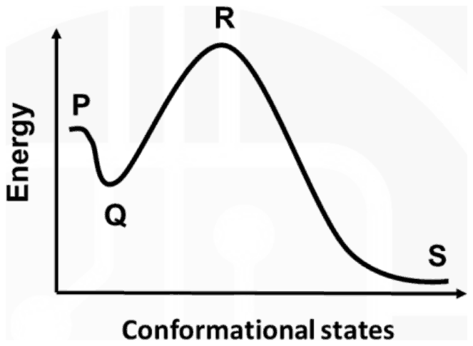
The following diagram represents energy states of different protein folding steps, which includes-

1. Folded
2. Transition
3. Molten globule
4. Unfolded

Energy against conformational states: the curve starts at P, dips to a shallow minimum Q, rises to a peak R and falls to the lowest level S

Find the correct match of folding steps (1 to 4) with the energy state (P to S) in the diagram.

Exam Year(s)

GATE Life Sciences - 2026,

Answer: A

The matching is 1-S, 2-R, 3-Q, 4-P.

Read the energy profile from left to right. P, the high starting level, is the unfolded chain (4). It drops into a shallow local minimum Q, the partly structured molten globule intermediate (3). The highest point R is the transition state (2) that has to be crossed. S, the lowest energy on the right, is the stable native folded protein (1).

The other options misplace a defining feature: the folded state must be the global minimum, so it cannot be R or P; the transition state must be the maximum, so it cannot be Q; and the molten globule is an intermediate, not the lowest state.

Please log in or register to report this question.

7.

During denaturation, which level of protein structure remains Intact?

Exam Year(s)

RSMSSB - 2025, RSMSSB Lab Technician - 2025,

Answer: A

Denaturation leaves the primary structure intact.

The primary structure is the sequence of amino acids joined by covalent peptide bonds. Denaturing agents such as heat, extremes of pH, urea or detergents break only the weaker hydrogen bonds, ionic bonds and hydrophobic interactions that fold the chain, so the sequence survives while the protein loses its shape and function.

The secondary structure (alpha helices and beta sheets) depends on hydrogen bonds, and the tertiary and quaternary structures depend on folding forces and subunit contacts. All three are disrupted by denaturation, which is why only the primary structure can be the answer.

Please log in or register to report this question.

8.

Which of the following amino acids among the following is preferred in a reverse turn ?

Exam Year(s)

MH SET - 2018, SET Exam - 2018,

Answer: A

The amino acid preferred in reverse turns is proline.

A reverse (beta) turn reverses the direction of the polypeptide chain over four residues, held by a hydrogen bond between the first and fourth. Proline's side chain is joined back to its own backbone nitrogen, fixing the phi angle near -60 degrees and readily allowing a cis peptide bond, which suits the second position of a turn. Glycine, which is very flexible, is the other residue common in turns.

Alanine and glutamic acid are strong helix formers, and histidine has no special preference for turns, so none of them is the residue typically found there.

Please log in or register to report this question.

9.

Which of the following is the most suitable mode to study protein conformational changes and to probe the location of active sites and coenzymes in fluorescence spectroscopy ?

Exam Year(s)

MH SET - 2019, SET Exam - 2019,

Answer: C

The key gives intrinsic fluorescence.

What could and could not be confirmed: searches confirmed that the intrinsic fluorescence of a protein, mainly its tryptophan residues (excitation near 280 nm, emission near 350 nm), changes in wavelength and intensity with the local environment, and so is the standard label-free way to follow conformational changes. They did not turn up a source that ties the phrase 'location of active sites and coenzymes' to intrinsic fluorescence rather than to extrinsic probes, so that half of the keyed answer rests on the University's key.

Intrinsic fluorescence needs no added dye, and a ligand or coenzyme bound near a tryptophan quenches or shifts its emission, which is how it can report on the neighbourhood of an active site. Quenching and quantum yield are measurements or processes within a fluorescence study, not a mode. Extrinsic fluorescence, which uses an attached probe, is the usual alternative and is the option a reader might defend; the key is the University's.

Please log in or register to report this question.

10.

The conformation of the polypeptide chain is determined by the torsion or rotation angles around Cα-N and Cα-C bonds of each amino acid participating in it. Which of the following pairs of amino acids is conformationally the most and the least restricted amino acids ?

Exam Year(s)

MH SET - 2019, SET Exam - 2019,

Answer: D

The most and least restricted amino acids are proline and glycine.

The backbone conformation is set by the angles phi (around Cα-N) and psi (around Cα-C). In proline the side chain loops back and bonds to the backbone nitrogen, forming a ring that fixes phi at about -60 degrees, so proline is the most restricted residue. Glycine has only a hydrogen as its side chain, so it meets the least steric hindrance and can take up many more phi-psi combinations than any other residue.

Option A gives the pair in the wrong order, and alanine, with its small methyl side chain, lies in between and is neither extreme.

Please log in or register to report this question.

Loading…

Other Subjects

Tags: Protein Structure and Function Questions and Answers || Protein Structure and Function MCQ Questions and Answers || Protein Structure and Function GK Questions and Answers || Protein Structure and Function GK MCQ Questions || Protein Structure and Function Multiple Choice Questions and Answers || Protein Structure and Function GK || GK on Protein Structure and Function || Biochemistry Questions and Answers || Biochemistry MCQ Questions and Answers || Biochemistry GK Questions and Answers || GK on Biochemistry